6
July
2022
The early-stage website of FEBS 2023 is launched
Guess the molecules in the second slider image of the homepage!
Wondering about the molecular representations in the homepage's image carousel? These are taken from PDB-101, the education portal of the Protein Data Bank, from the illustrations in 'Molecule of the Month' by David S. Goodsell.
For non-structural biologists, here are hints to identify them, from left to right:
- pre-mRNA proccesing
- a way to study membrane proteins
- a key moment in gene expression
- mitochondrial energy transfer
Answers:
- 'Yeast spliceosome captured after the splicing reaction has occurred, with the intron "lariat" shown in yellow. Proteins are in blue and green, and spliceosome RNA molecules are in magenta and pink' (https://pdb101.rcsb.org/motm/245).
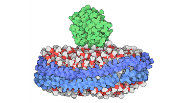
-
'Membrane-binding domain of MT1-MMP (green) bound to a nanodisc' (https://pdb101.rcsb.org/motm/237). A nanodisc is a tiny disc of lipids that allows structural studies of a membrane-bound protein; the metalloproteinase MT1-MMP is implicated in cell migration through extracellular matrix, including in metastasis.
-
'RNA polymerase (blue) stalled while unwinding a nucleosome (orange, with DNA in red). Several elongation factors are in green, and a little piece of the transcribed RNA is seen poking out in magenta' (https://pdb101.rcsb.org/motm/241).
-
ATP synthase (https://pdb101.rcsb.org/motm/72). The membrane-embedded F0 motor (top blue region) uses the power from a proton gradient to force the F1 motor (lower pink region) to generate ATP.
Images by David S. Goodsell and RCSB PDB (CC-BY-4.0 license).